

First-time Ultrasound

What is an ultrasound, actually?
Have you ever had another mama show you a picture of an ultrasound (or "echo") of her baby when it was still in her belly?
An ultrasound not only makes everyone excited, but it is also an exam that reveals a lot of important things for the mama and the baby.
How is an ultrasound done?
An ultrasound is an examination method that is performed beginning with the first visit.
It checks the health of the baby inside and how it's growing by obtaining images of the maternal-fetal condition.
Ultrasound technology is very advanced these days, and there are various types available with different ways of placing a probe and different ways of displaying the images.
How an ultrasound works
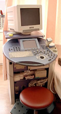
a Machine for ultrasound
An ultrasound is often compared to a fish-finder machine.
When ultrasonic waves, which are high-frequency, pass through liquid, they have the characteristic of reflecting at the edges of organs and tissues, etc.
Applying this in the ocean led to the fish-finder machine, and applying it in a mama's belly filled with amniotic fluid led to ultrasounds for exam use during pregnancy.
It can display the reflected waves as images, so you can check things visually on the monitor and sometimes even receive a print-out or video recording, depending on the hospital.
Unlike X-rays, ultrasounds have not been reported to have adverse effects on pregnant women.
They can be considered a very safe examination method.
Types by a probe position
Transvaginal method
a photo of ultrasound by
a transvaginal method
In this method, a rod-like ultrasonic wave-emitting device called a transvaginal probe is inserted into the vagina to perform the examination.
This method is used until about the 11th or 12th week of pregnancy, so it is performed primarily for the ultrasound at the first visit. By inserting the probe into the vagina, the uterus can be observed from a position closer to the baby inside the womb, enabling a precise image to be viewed.
The doctor checks not only how the baby is doing but also the health condition of the mama's uterus and ovaries.
If you relax during the exam you won't feel pain, but if you get nervous and tense up, it may hurt when the probe is inserted. If that happens, be straightforward and tell the doctor.
The probe is sterilized after each use and a special condom is placed on it, so cleanliness is thoroughly taken care of.
Transabdominal method
a photo of ultrasound by
a transvaginal method
The ultrasounds performed from the second trimester of pregnancy use a method that probes from the outside of the belly.
The doctor rubs a gel on the belly that helps the ultrasonic waves pass through the skin, and simply slides the probe around little by little on the belly. The gel may feel cold, but there is no pain. However, you may be asked to change your position or breathing in order to work around the baby's position or movements, and moving as instructed can be difficult in the third term of the pregnancy.
The gel is of course harmless, and it comes off fine by simply wiping it with a cloth after the exam.
Types by exam methods
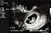
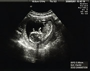
Ultrasound tomography (2D)
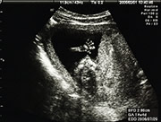
a photo by 2D ultrasound
This method displays the information obtained by the ultrasonic waves' reflections as a 2-dimensional (flat surface) image, and it is the exam method most often used.
It can be done either transvaginally or transabdominally.
Color Doppler method *Transabdominal method
This ultrasound method lets doctors measure the amount of blood flowing and its speed, and it also reveals things like the number of blood vessels in the baby's umbilical cord and abnormalities in the shape of the baby's heart.
3D method *Transabdominal method
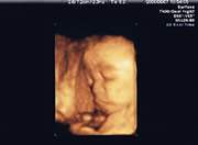
a photo by 3D ultrasound
With this method, you can observe the baby inside as a 3-dimensional, realistic shape, just as if you were peeking at the baby through a window.
4D method *Transabdominal method
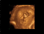
a photo by 4D ultrasound
This method adds the passage of time to the 3D method and record it in a moving image.
You can see the baby moving comfortably and softly in the amniotic fluid, just as if it is taking a space walk.
To check the baby's growth and evaluate the condition of the mama's uterus and ovaries, a 2D method works just fine. Consequently, there are not many maternity hospitals that offer 3D and 4D methods yet. Even if they have 3D and 4D available, in many places they are not necessary exams but are offered only upon request.
Ultrasound frequency
While it depends on the medical facility, since ultrasound exams cost money, some maternity hospitals may do them only 2 or 3 times as needed, whereas others may do one at every regular visit.
What can you find out from an ultrasound?
In a 3D or 4D ultrasound, the baby is very real-looking.
It's hard to believe it's an image from inside the uterus.
But what is looking at a baby inside mama's body like this intended to help us find out?
Checking mommy's health condition
First, it lets the doctor check the condition of the mama's uterus and ovaries. During the initial visit, it is an essential exam for checking if the pregnancy has any problems, such as whether it is an ectopic pregnancy or whether the ovaries have any abnormalities, etc., in order to make important decisions.
Once the pregnancy has progressed, it is useful for evaluating things like a placenta previa.
Knowing how the baby is growing
The primary goal is to check whether the baby is growing properly. Not only can it find out if there are twins or multiple fetuses, or if the baby is breech, but depending on the baby's position, it can also make it possible to measure the length or size of various parts. The doctor will measure the length from the baby's head to his bottom, a measurement called the "crown-rump length" that is used to determine the due date, as well as other detailed measurements such as the diameter of the baby's head, the length of the thigh bone, and the size around the belly.
At around the 24th week of pregnancy, the distinction between male and female will appear. However, since the exam is not done for the purpose of evaluating the gender, some maternal hospitals won't tell you that information.
Knowing the baby's health condition
Because a color Doppler method shows the baby's blood flow, it can also show the health condition of individual body parts and how the umbilical cord is doing, and it may find abnormalities in the heart, for example.
Also, based on the amount of amniotic fluid, the position of the placenta, and how the umbilical cord is doing, it can find information ahead of time that could bring disadvantages at the time of birth. But because it is after all a visual exam and differs from a scientific one, not all conditions can be detected with this exam.
More than what you "find out," it's what you "feel"
Being able to "see" the baby inside the belly with an ultrasound is a very magical thing. This is the first experience where mommy gets to "meet" her baby. It is very useful in bringing about a maternal awakening as well. If you can get a picture, be sure and show it to your family right away.
Can I get a picture?
Many people have pictures from when they had ultrasounds performed.
So some people may look forward to the exams.
Some can even get videos as well.
Most obstetrics and gynecology (OB/GYN) departments will give you a picture as a keepsake, but the exam is done strictly for evaluation purposes.
It is an exam that doctors perform at their own medical discretion, so ask your primary doctor.
If you want a picture...
If you want an ultrasound picture, the best idea is to check whether you can get one or not when choosing an OB/GYN. However, if you have already chosen an OB/GYN without knowing about all that, there are lots of medical facilities that will photograph an ultrasound image for you even if you are not a regular patient there. Just search for one on the Internet, etc.
Can I get the picture for free?
That seems to depend on the medical facility as well. If it's an ordinary 2D image, there are lots of places that will give you a print-out as a keepsake in good faith. But a picture of a 3D image or a video will usually cost at least three thousand pesos.
update : 19.09.2017
Our favorites feature uses your browser's cookies. To use this feature, please enable cookies. If you are using Safari on your iPhone or iPad, please turn off the Private Browsing Mode. If you clear the cookie, you also clear the Favorite that you chose.